Project ongoing · Synthetic biology
BioBrick
We design pieces of DNA to grow more faithful organoids in the laboratory.
- the scar left between two parts
- 8 bases
- cut and join the standard parts
- 4 enzymes
- the pSB1C3 plasmid, to ship the parts
- 2070 bp
- teams competing at iGEM, in 2025
- 412
For people who come from biology, biotechnology, bioinformatics
The BioBrick standard
A BioBrick is a piece of DNA with a precise function, for example switching a gene on, and with the same ends for everybody. The standard is called RFC 10: Tom Knight wrote it at MIT in 2003, and it is the most used one in synthetic biology.
Every part has the same prefix upstream and the same suffix downstream, with the sites of four enzymes that cut DNA. By joining two parts you get a new part, with the same prefix and the same suffix, which is reused in the same way, endlessly.
Between the two parts a scar of 8 bases is left, TACTAGAG: it appears where the end cut by SpeI binds the end cut by XbaI, and neither enzyme recognises it any more. The limit is well known: in fusion proteins the scar shifts the reading of the genetic code, and there other standards are used.
prefix    GAATTCGCGGCCGCTTCTAGAG
          GAATTC                    EcoRI
                GCGGCCGC            NotI
                         TCTAGA     XbaI

part      ... the sequence with the function ...

suffix    TACTAGTAGCGGCCGCTGCAG
           ACTAGT                  SpeI
                  GCGGCCGC         NotI
                         CTGCAG    PstI

scar TACTAGAG 8 bases between two assembled parts| EcoRI | GAATTC | cuts G^AATTC, in the prefix |
|---|---|---|
| XbaI | TCTAGA | cuts T^CTAGA, in the prefix |
| SpeI | ACTAGT | cuts A^CTAGT, in the suffix |
| PstI | CTGCAG | cuts CTGCA^G, in the suffix |
| NotI | GCGGCCGC | in the prefix and in the suffix |
A transcription unit
To make a cell produce a protein you need four parts in a row on the DNA. The drawing uses the symbols of the SBOL Visual standard, the same ones in every laboratory.
- 01
Promoter
A bent arrow: this is where the reading of the gene starts. It decides when the gene is on and how much.
- 02
RBS
A dome on the DNA line: the point where the ribosome hooks on and builds the protein.
- 03
Coding sequence
A solid arrow: the gene itself, with the instructions for the protein.
- 04
Terminator
A T on the line: here the reading stops.
How two parts are assembled
The method recommended by the iGEM Registry is called 3A assembly: three different antibiotics select the right constructs by themselves, with no gel and no PCR.
- 1The left part is cut with EcoRI and SpeI.
- 2The right part is cut with XbaI and PstI.
- 3The destination plasmid, which resists a third antibiotic, is cut with EcoRI and PstI.
- 4Everything is ligated together: the SpeI end joins the XbaI end and the scar appears.
- 5It is grown with the third antibiotic: the correct constructs survive, in theory about 97%.
| Length | 2070 base pairs | |
|---|---|---|
| Resistance | chloramphenicol | |
| Origin of replication | pMB1 | derived from pUC19 |
| Copies per cell | from 100 to 300 | |
| Terminators | on both sides of the part | they isolate it from the rest of the plasmid |
The double helix, to scale
The drawing follows the measurements of B-DNA, the most common form in cells. The two strands are not opposite each other: this is why a wide groove and a narrow one open between them.
| Base pairs per turn | 10,5 | |
|---|---|---|
| Rotation per base pair | 34,3° | |
| Rise per base pair | 0.332 nm | |
| Helix pitch | 3.32 nm | one full turn |
| Diameter | 2 nm | |
| Major groove | 2.2 nm | |
| Minor groove | 1.2 nm | |
| Handedness | right-handed |
The numbers in the chart
| Piece of DNA | Length |
|---|---|
| A scar (8 bases) | 2.7 nm |
| A typical part (1000 bases) | 332 nm |
| The pSB1C3 plasmid (2070 bases) | 687 nm |
Organoids
An organoid is a group of cells from an organ that grows from stem cells and organises itself, as in the body. The definition is by Lancaster and Knoblich, in Science, in 2014.
An intestinal organoid is a layer of cells around a cavity, the lumen. Buds grow outwards that copy the crypts of the intestine, with the stem cells and the Paneth cells at the bottom. Old cells fall into the lumen, as in the real intestine.
It does not grow beyond a few millimetres because it has no blood vessels: oxygen only reaches 100 to 200 micrometres from the surface, and further in the cells die.
The numbers in the chart
| Measure | Size |
|---|---|
| Oxygen without vessels (how far it enters the tissue) | from 100 to 200 µm |
| Intestinal organoid (typical diameter) | from 200 to 500 µm |
| Brain organoid (the largest ones) | up to 4 mm |
They are used to test drugs. In the reference study, with patients who had gastrointestinal tumours, the response of the organoids predicted the response of the patients. On 10 April 2025 the American FDA announced a plan to reduce animal testing for some drugs, replacing it with organoids among other things, over a period of 3 to 5 years.
The numbers in the chart
| Measure | Value |
|---|---|
| Sensitivity (the drugs that work) | 100 % |
| Specificity (the drugs that do not work) | 93 % |
| Positive predictive value | 88 % |
| Negative predictive value | 100 % |
Circuits inside cells
A genetic logic gate is a small network of genes. The input is a molecule, for example a drug or a toxin, that switches a promoter on or off; the output is a protein. With promoters activated or repressed by different proteins you build AND, OR and NOT, as in a computer.
In 2011 colonies of Escherichia coli worked as NOR gates connected by chemical signals. In 2016 the Cello compiler translated the description of a logic circuit directly into a DNA sequence. Our goal is to bring this idea inside an organoid: a circuit that recognises a drug or a toxin and switches on a signal that can be measured.
iGEM
iGEM is the international synthetic biology competition. It was born as a course at MIT in 2003; the first competition, in 2004, had five teams. The parts made by the teams end up in the Registry, a public collection with more than 70,000 documented parts.
Since 2022 the final, the Grand Jamboree, has been held in Paris. We are aiming at the 2027 edition, whose date is not published yet.
The numbers in the chart
| Year | Teams |
|---|---|
| 2023 (44 countries) | 393 teams |
| 2024 (41 countries) | 397 teams |
| 2025 (44 countries) | 412 teams |
Where we are
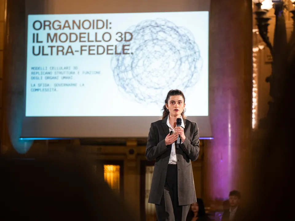
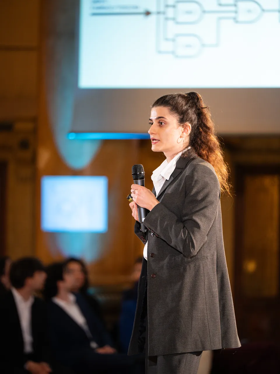
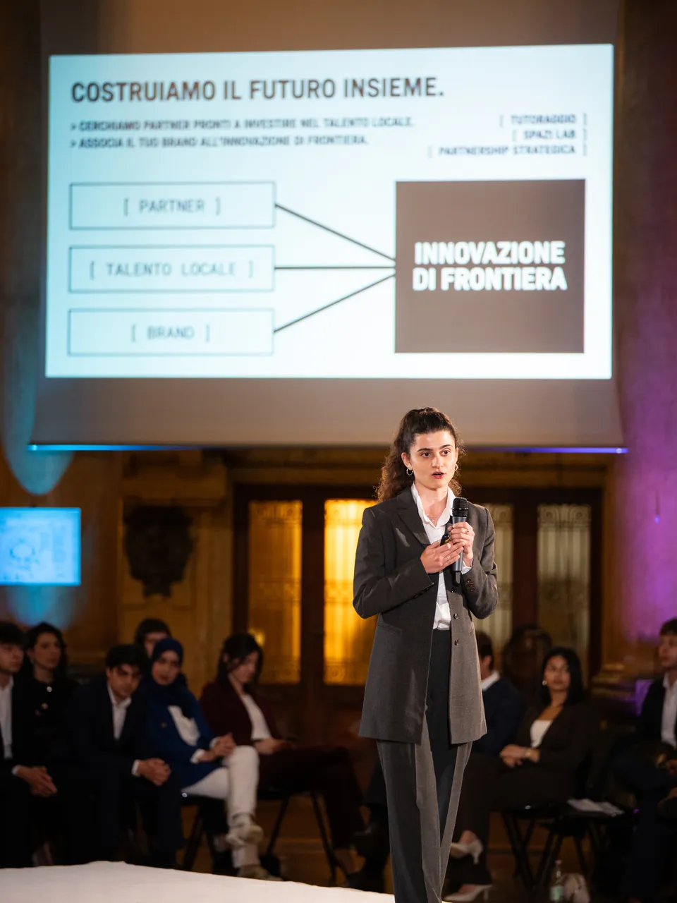
- April 2026We presented the project at Palazzo della Borsa, in front of universities, companies and institutions.
- TodayThe team brings together molecular biology, bioinformatics, computer science and doctoral research.
- 2027We are aiming at iGEM, the international synthetic biology competition: the Grand Jamboree, held in Paris since 2022.
Photos
People on the project
Sources
- iGEM Registry, BioBrick RFC 10 standard and 3A assembly: parts.igem.org.
- SBOL Visual 3.0: sbolstandard.org.
- Lancaster and Knoblich, Science, 2014; Sato and colleagues, Nature, 2009.
- Vlachogiannis and colleagues, Science, 2018.
- FDA, announcement of 10 April 2025 on animal testing.
- Tamsir, Tabor and Voigt, Nature, 2011; Nielsen and colleagues, Science, 2016.
- iGEM, past editions: competition.igem.org.
- Presentation at the Deep-Tech Showcase, Palazzo della Borsa, 21 April 2026.
Do you want to work on it?
You do not need experience and you do not need a CV. Write to us: we invite you to the next meeting, where you meet the team.